 Whatsapp
 Call Us


Ultrasonography
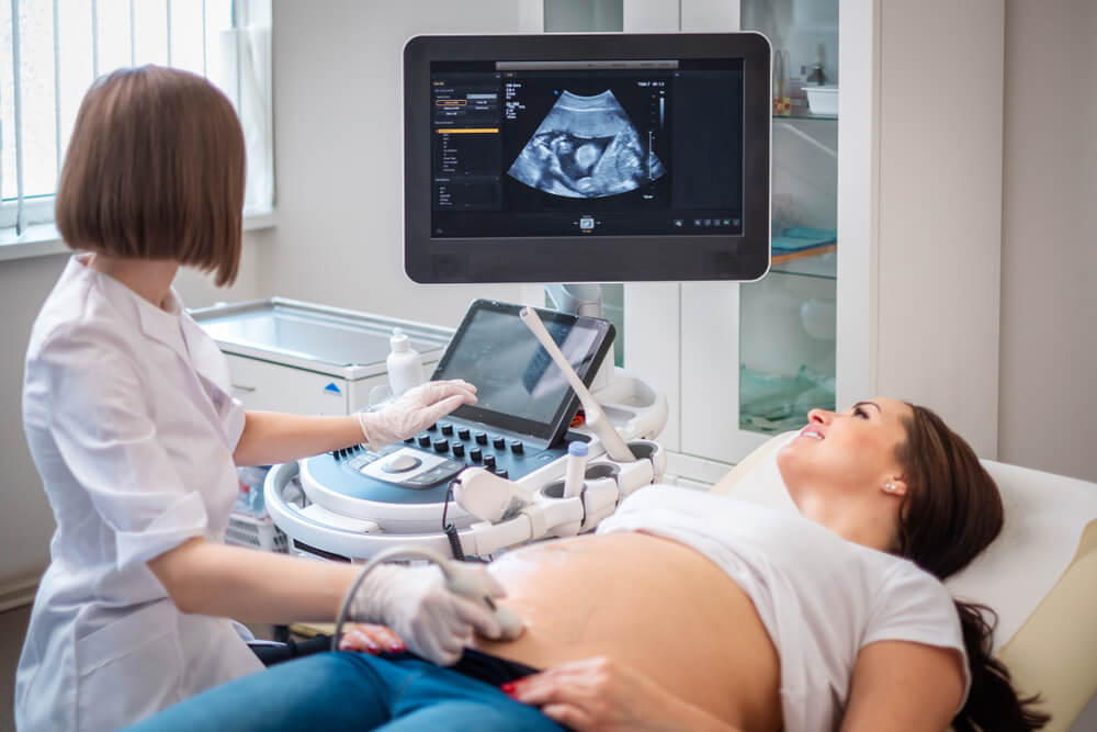
What is Ultrasound?
Ultrasonography is a diagnostic method in which sound waves are used in a detailed examination of the internal contours of the body. It is often preferred as a technology that provides benefits in diagnosing many diseases and responding to treatment at an early stage.
Many diseases in people are caused by internal organs and physical examinations may be insufficient in some cases. For this reason, ultrasound imaging is often used in the medical world to provide an opportunity to study the causes of diseases in more detail. Ultrasonography imaging, which may be needed in every field, is an important diagnostic system applied especially in the detailed examination of internal organs.
It is an imaging technology that provides benefits for doctors to accurately diagnose diseases and start treatment at an early stage. Ultrasound technology, which is quite safe because it does not emit radiation, helps to make clear diagnoses without noticing a woman or a man.
As Safir laboratory, ultrasound imaging is performed in the most practical way by using advanced devices in our center. Our specialist physicians can examine the images of our patients in detail as desired while providing services in this field. With the help of our devices using the latest technology, results can be obtained in a short time without waiting and the treatment process can be started.
It is an imaging device that provides a great advantage since it does not have any harm to the baby and mother during pregnancy due to the fact that it is not a radiation emitting system. It is an important aid for the early diagnosis of many health problems such as trauma, internal tissue injuries, where results can be obtained in a short time.
Ultrasound shots are usually completed in a period of 15 minutes to 40 minutes. Dec. The change of periods depends on which ultrasound device people need and what their complaints are.
While the detailed shots take longer, the images used to diagnose other diseases may take shorter.
Ultrasound scans, which are completely completed in periods that differ depending on the patient and the physician’s expertise, are often used in the diagnosis of most diseases. It is a technology that benefits the early initiation of treatment and the recovery of the patient and has an important place in the field of medicine.
Field of Use of Ultrasonography
The most widely used ultrasonography devices in the medical field are an important device for observing areas of the body that cannot be seen with the eye and cannot be reached endoscopically. This diagnostic method, which is used in the diagnosis of many diseases, is often preferred so that changes in the body can be observed and any adverse situation that may occur can be intervened at an early time.
Ultrasound imaging, which is often used in pregnancy follow-up due to its harmlessness in the organs located in the abdominal cavity, offers patients the possibility of treatment in many areas. The fact that the devices are of high quality and the images are examined by specialists benefits from starting the treatment correctly.
For this reason, while the imaging of our patients is performed clearly by using the devices manufactured with the latest technology in our center as safir laboratory, the results and the process are carried out by expert people.
Other areas where the imaging system is used in pregnant women infant and maternal health screenings, heart health problems, infections, to investigate the possibility of breast tumor and a surrounding, Nov diseases, problems in the genital area, takes place in the display of fields, such as gallstones.
These health problems can bring serious problems if they are not treated and a definitive diagnosis is not made.
Ultrasonography, which has a very important place in the medical field, has an important place today thanks to the developing technology. It can be used because it is a safe procedure in all areas of the body.
Advantages of the Ultrasound Method
The ultrasonography system, which provides benefit in the examination of anomalies in all internal organs located in the body, is a frequently preferred diagnostic technique in medicine due to its harmlessness.
Thanks to this diagnostic technique, it provides the possibility of treatment at an early stage, allowing diseases to recover without making progress. There is no harm to patients due to the absence of radiation emission and it is often preferred for pregnancy scans.
The patient can return to his normal life in a short time without experiencing any pain or pain sensations during the shots.
It is not necessary to stay in the hospital without a disruption that will affect his life and health. It is an easy-to-apply method and the structure of organs and blood vessels can be safely examined in detail.
Since it is a method that has no harm or side effects, it is a necessary method for examining the health of the baby and mother during pregnancy. There are different types of ultrasonography systems that provide imaging in many parts of the body according to the areas to be used. It is very important that the correct varieties of this device are used according to the areas used and that it is managed by an expert person.
In order for clear images to be taken, it is important to act according to the different types of the disease and the region to be examined, the correct lying position, the recommendations of the specialist physician. Thus, it becomes easier to make the correct diagnosis and take clear images.
Pregnancy Follow-Up with Ultrasonography
The examination of maternal and infant health during pregnancy is of great importance. During this period, it is necessary to examine all the details by specialist physicians without harming the health of the baby and mother, and to conduct screening. The ultrasound device is one of the healthiest screening techniques used for pregnancy monitoring and is the most commonly used examination method during this period.
This screening method used in pregnancy follow-up is extremely safe both for the baby and for health. It is often used as a screening technology requested by a specialist physician during certain periods of pregnancy. Examination of the fetus during pregnancy, the development of the baby, heartbeat, formation of organs, weight and height can be examined in detail, in case of any abnormalities, the treatment process deemed necessary by specialist physicians is initiated.
Pregnancy problems are generally 20-24. It can be diagnosed in weeks. Images are obtained in 3 Decimals or 4 dimensionsby using the type of ultrasound required between these weeks. According to the results obtained in the images, the pregnancy is terminated or the intervention that needs to be performed can be started at an early stage.
Ultrasound devices used to obtain clear images in infants, to examine limbs, to diagnose structural diseases early are used in the highest quality in our safir laboratory center. Due to the fact that the images taken with high-quality devices are clearer, specialists who offer treatment opportunities at an early stage are serving in our center.
Colored ultrasound and 3-4 dimensional ultrasound shots called detailed during pregnancy are generally preferred. A detailed examination of the health of the mother and baby is also a kind of ultrasound, which is necessary for easy management of the whole process. In case of any negativity in pregnancy scans, different examinations are performed to ensure that the process continues in a healthy way.
Ultrasound shots, which are important for the health of both the mother and the baby, should definitely be performed by a specialist. Most of the time, perinatology specialists are involved in detailed shootings. In this way, healthy images are obtained without skipping all the details. It is important not to neglect ultrasound shots and to take shots during the periods recommended by specialist physicians in terms of maternal and infant health.
Ultrasound Preparation
Ultrasound, which is used for many health screenings and gives successful results in maternal and infant health screenings, especially during pregnancy, has an important place in the field of medicine.
Getting counseling and informing the patient before the ultrasound is also a very important issue for the patient’s comfort. In this way, the patient leaves the procedure in a short time, allowing the results to come out clearly.
Prior to ultrasound imaging, patients are informed by specialist physicians and qualified specialists. In these informations, it should be ensured that the patient is immobile and the areas where his complaints are located are thoroughly determined.
The correct determination of the complaints between the patient and the specialist physician during the examination allows the ultrasound examinations to be performed Decisively.
After the correct choice of ultrasound devices, the shooting is completed in a short time. There is no need for any preliminary preparation stage, except for the patient’s examination.
Different preliminary preparations may be required depending on the type of ultrasound imaging. Especially if shots are to be taken in the abdominal region, if the patient is asked to starve, and if an examination is to be performed in the bladder area, it is absolutely necessary that the patient is stuck in the urine. For this reason, preliminary preparation may be required depending on the area to be shot.
It is necessary to apply gel to the area to be imaged in order to get clearer pictures of the area to be imaged. The gel does not cause any side effects and helps to transmit radio waves by lubricating it so that the area can be viewed more clearly.
After these preliminary preparations are completed, the prop device is moved around the designated areas and the necessary images are transferred to the monitor. The images transferred to the monitor are reported and sent to specialist physicians by the radiology specialist. After the examination of the images, the necessary treatment process begins and an intervention takes place quickly for the patient to recover.
Diagnosis of Diseases by Ultrasound
With the use of ultrasound techniques and different devices, the diagnosis of many diseases can be made easily. Imaging systems that are frequently used today are technologies that provide benefits for the diagnosis of diseases in the medical field and for early diagnosis.
Transvaginal, colored doopler and many other different types of ultrasonography are used to diagnose diseases. After the controls of the specialist physicians, he informs the patient about which ultrasound device should be used and which areas should be examined.
After this information, the patient is taken for ultrasound accompanied by a radiology specialist. Which device is preferred is important in the diagnosis of diseases. That is why it is important to correctly implement the correct devices. The correct and clear appearance of the results depends on the correct ultrasound scan and allows the person to start the treatment process at an early stage.
As Safir laboratory, we ensure that the results are obtained as soon as possible by using all ultrasonography devices successfully in our center.
We are working quickly for the successful progress of the treatment process by making quality shots in our center where expert radiologists serve. By prioritizing the comfort of our patients, all our devices have been provided to provide the Jul-test quality service.
Ultrasound Prices 2026
It is necessary to decide and determine which imaging device will be used for ultrasound imaging and which region should be examined in advance. While determining whether the ultrasound will be detailed or not, information on the prices is taken at the same time.
Details such as the type of ultrasound, its details, the region where the imaging will be performed, and the doctor’s specialty make changes in prices. In public hospitals, procedures are performed free of charge upon the request of specialist physicians, while in private hospitals, procedures are performed on a paid basis.
Centers where the shots are not made correctly should not be preferred by being deceived into the fact that the fees are appropriate. It is absolutely important to conduct research beforehand. Performing the shots in appropriate conditions also causes the treatment process to be performed in a healthy way. For this reason, it is necessary to evaluate the reports by specialist physicians while the shots are performed correctly by a specialist person.
We provide services to our patients at the most affordable prices while using the highest quality devices in our Safir laboratory health center. Accompanied by our expert physician staff, we are beneficial to ensure your recovery opportunity by starting your treatment process quickly.
By doing research, you can make an appointment at our center for the most affordable ultrasound prices and get information from our expert friendly staff. You can contact us immediately to get information about ultrasound prices 2026.