 Whatsapp
 Call Us


Knee MR
What is the Knee MR?
Knee MR is an imaging technique used for knee problems. It is possible that various problems occur in different body parts of people. One of these regions comes to the fore as the knee region. Diagnosis should be made in order to eliminate the problems that occur in the knee. You can get services from our health center for this diagnosis and have an MRI.
In case you have knee complaints, our doctors may ask you to have an emar. One of the most widely used diagnostic methods in the field of health is to use EMAR. You can have problems related to your knee detected and treated by having a knee reconstruction at our Safir Lab health center. Magnetic resonance imaging is extremely important in the examination of the skeletal system, organs, tissues.
If you experience pain and problems in your knee, it is useful to care about them. Otherwise, it is possible that the problem will progress and become a more serious problem. Especially if you feel a lot of pain and pressure in your knee area, you should definitely have an MRI. After MRI, our specialist doctors make the necessary assessment and diagnose if you have a disease.
After the diagnosis, the treatment process is started in a short time. Early diagnosis is very important for easy treatment. Late diagnosis may cause the treatment process to be prolonged. People who want to get precise and clear results can get services from our health center, which provides services with specialist physicians and medical personnel in the field.
There may be more than one factor that causes you to experience knee pain. Accidents, blows, sports injuries, genetic problems and similar factors are among the most important of them. Our doctors may order an MRI (Magnetic Resonance Imaging) of your knee. MRI is used to obtain images of internal body structures or bones and tissues.
MRI of the knee allows doctors to examine the anatomy of the knee to rule out any structural abnormalities. An MRI of the knee (magnetic resonance imaging) uses radio waves and a magnetic field to create images of the internal and surrounding anatomy of the knee. Our doctors may recommend a knee mr in a variety of different situations.
How is the Knee MR Taken?
The most commonly used method in case of any problems with your knee is imaging techniques. Within the scope of these techniques, it is possible to determine the cause of knee problems. It is an extremely fast performed operation. We take MRI in our health center in a short time and detect your knee problems.
Before MRI, our medical personnel inform you about the necessary issues. With the information we provide, we explain that MRI is not a situation to be afraid or worried about. After providing the necessary information to the patient, we obtain information about the medical history. If there are no obstacles in the patient’s medical history, a knee emar extraction is performed.
If you have any metal items before the MRI scan, we would like you to leave them. MRI should not be performed with metal products. Apart from this, you do not need to do any October preparation. Imaging procedures, in general, do not require special preparation. You need to leave jewelry, keys, coins, accessories and similar things. Then we start the procedure by taking our patients to the emar room. The Emar shooting will be completed in a short time.
With Knee MRI, special scanning equipment takes images of your knee. The technologist can use a special coil that acts as an antenna to help shorten scanning times and produce higher quality images by focusing the power of magnetic and radio waves on your array.
The MRI software transfers the images to a HIPAA-compliant computer. A radiologist then examines the images on a high-resolution monitor to assess the soft tissue of the joint.
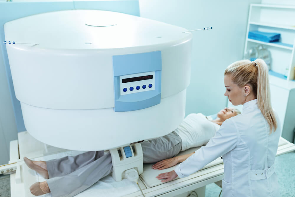
With a traditional MRI machine, you lie down on a padded table, which will move to the scanner so that the technologist can take images of your knee. The technician will communicate with you throughout the process and check how you are doing. It is important to be as still as possible during this part of the examination so that we can capture the highest quality images for the radiology doctor to interpret.
There are also standing MRI machines and upright open MRI machines for patients with claustrophobia, who cannot lie down comfortably, or whose dimensions prevent them from being evaluated in a conventional MRI scanner. During these scans, you can stand or sit unhindered facing forward
The technologist will help you lie down comfortably on a cushioned table, offer you earplugs or headphones and a blanket to make you comfortable. A standard knee MRI takes about 30 minutes and there is no pain during the examination.
Depending on your doctor’s instructions, an MRI contrast (a special dye that helps to highlight your anatomy) may be required. The contrast will be administered through an IV placed in your hand or arm before the exam. Contrast helps to emphasize the anatomy of your knee in the captured images.
Why is Knee MR Requested?
Our specialist doctors decide whether an MRI is needed for the knee. In general, in case of a problem related to your knee, our specialist doctors may request an MRI. It is requested for pain and complaints that occur in the knee area and around it in general. It is important to have an MRI to find out why the patient’s complaints are caused.
Thanks to MRI, it can be easily determined what the knee problem is and the treatment method is determined by our specialist physicians. Physical control and treatments for knee pain and problems do not work definitively in some cases. For the correct diagnosis and treatment, imaging techniques come to the fore.
With Emar withdrawal, we make the correct diagnosis for the patient’s knee problem. It is normal for problems to arise in your knee due to incorrect movements, accidents and similar situations. Many people may not care, thinking that the problem they are experiencing is temporary. However, with the further progression of the situation, big problems may arise. For this reason, we recommend that you have an MRI if you are experiencing regular problems in your knee or any other area. The area where there is a problem with the knee emar is displayed in detail and clearly. In this direction, the source of the problem is determined and the treatment process is started.
MRI imaging for the knee is performed for specific purposes:
- Check the cause of unexplained knee pain or the knee sticking out for no reason.
- Find problems in the knee joint such as arthritis , bone tumors or infection, or damaged cartilage, meniscus , ligaments or tendons.
- Find out if knee arthroscopy is required.
MRI can also find a bone fracture when X-rays and other tests do not give a clear answer. MRI is performed more often than other tests to check for certain bone and joint problems.
MRI of the knee creates more detailed images than a traditional X-ray and is safer because it does not use radiation. Healthcare providers use knee MRI techniques to assess and diagnose a range of problems:
- Damage to the cartilage, ligaments, tendons or meniscus of the knee
- Sports injuries
- Knee pain
- Fluid buildup in your knee
- Infections
- Problems with a number of movements
- Prepare for knee surgery
- Follow-up after knee surgeries
Where is the Knee MR Taken?
The knee is one of the areas where people experience the most pain and similar problems. In this direction, it is possible that you often encounter knee problems. Even if sometimes this situation may be insignificant, sometimes it can be extremely serious. It will be very useful to have an MRI together with physical therapy or after physical therapy.
By having an EMAR taken at our health center, you can find out exactly why the problem in your knee is caused. It is important that you definitely have a knee reconstruction to prevent possible health problems that may occur in the future.
You can have an MRI for knee problems that are not visible to the eye and are caused by the inner part of the body. As Safir Lab, we provide services in this regard in our fully equipped center.
The imaging process using magnetic resonance is performed by a machine that uses radio waves and a magnetic field. This machine provides detailed display of the index. Muscles, ligaments, cartilage and other joint November structures are usually best seen with MRI. In most cases, MRI gives information about structures in the body that are also not visible by X-ray, ultrasound or CT scan.
For the MRI test, you are placed inside the magnet so that your knee is inside the strong magnetic field. MRI can find changes in the structure of organs or other tissues. It can also find tissue damage or disease, such as an infection or a tumor.
The images obtained as part of the MRI scan are sent to computers. At the same time, there is an opportunity to store these images on a computer. It can be used again later if needed or if it needs to be examined by a doctor.
Some Information About Knee MR
The emar process is performed by using radio waves in magnetic fields. The emar shot that will be performed does not have any negative effects for the patient. It also does not cause a risk to the person’s health status in the future.
If emar is required for knee problems, the procedure is performed in a short time. Depending on the situation, it is possible to extend it for up to half an hour. The device used for knee emar is open on three sides. In this way, there is no situation that will cause people with a condition such as claustrophobia to be afraid. The release time of the Emar results is also quite short.
The results are presented in a report and evaluated by our specialist physicians. After the evaluation, the appropriate treatment process is planned for your current problems. It is very easy to eliminate the discomfort with early diagnosis and treatment of knee problems.
To prepare for a knee MRI, it is useful to talk to your doctor about what to expect. Tell your doctor if there is any metal on or inside your body, including ear implants, clips for brain aneurysms, and/or a pacemaker.
At the beginning of the MRI, a technologist will ask you to remove items such as jewelry, metal zippers, and metal hair items. Also, remember not to wear makeup, as it can disrupt the viewing process. MRI examinations do not require special preparation. Unless your doctor recommends otherwise, eat and drink normally and follow your prescribed medication dose.
You will not have pain due to the magnetic field or radio waves used for the MRI test. However, you may be tired or sore from lying in the same position for a long time.
If a contrast agent is used, you may feel some coolness when it is placed in your IV.
In rare cases, you may feel:
- Those who have metal fillings in their teeth may experience a tingling sensation.
- The temperature in the controlled area. This is normal. Tell the technologist if you have nausea, vomiting, headache, dizziness, pain, burning, or breathing problems.
Knee MR Prices 2025
Imaging techniques are used to diagnose diseases all over the world. These techniques are quite effective in making the correct diagnosis of diseases. At the same time, it is also of great importance in terms of early treatment processes. It usually provides an advantage by completing the process in a short time and the results come out quickly.
MRI comes to the fore as a painless and side-effect-free imaging method. MRI can be used to control patients smoothly. As Safir Lab, we perform MRI scans for various parts of your body within the scope of the services we offer. People who are experiencing knee problems or ailments can have a knee reconstruction by getting services from our health center. You can contact us immediately to get information about knee mr prices 2025.