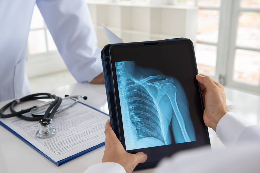
Digital X-Rays

What is a Digital X-ray?
Digital X-ray is the most widely used imaging technique for imaging internal organs. Image shooting is carried out with the use of various contrast agents. With this imaging, it is possible to detect many health problems of the person.
As Safir Lab, we use imaging technique in the examination of esophagus and urinary tract or in the examination of stomach and kidneys. It is also a widely preferred method in the examination of small and large intestinal canals. Digital X-ray performed with the use of X-rays is a method that does not cause any harm to the patient. We transfer the images we have obtained to the computer screen and conduct a more detailed investigation. It is an extremely effective imaging technique for detecting diseases. At the same time, it is possible to apply this technique without using a contrast agent.
X-ray imaging (radiography) is still the most widely used technique in radiology. To create a radiograph, a part of the body is exposed to a very small amount of X-rays. X-rays pass through tissues and hit a film or detector to form an image. X-rays are safe when used appropriately by radiologists and technologists who are specially trained to minimize exposure.
After the radiography is taken, there is no radiation left. Digital radiography (digital X-ray) is a form of X-ray imaging in which digital X-ray sensors are used instead of traditional photographic film. The advantages include increased efficiency by eliminating the need for chemical processing and the ability to transfer and enhance images digitally.
In addition, less radiation can be used to produce a contrast-enhanced image similar to conventional radiography. X-rays can be used to image all parts of the body and are most commonly used to look for fractures. They are also widely used for examining the chest, abdomen and superficial soft tissues. X-rays can identify many different conditions and are usually a quick and easy method for your doctor to diagnose.
A digital X-ray or digital radiography, as in traditional X-rays, is a modern type of X-ray that uses digital receivers instead of photographic film. The captured image is instantly converted into digital data and becomes ready for examination in seconds.
Almost everyone who has visited the dentist or broken a bone has had an X-ray in the past. Digital x-rays are the next step in the evolution of X-ray technology. They provide all the ease and convenience of traditional X-rays to recognize problems with bones, teeth, organs and other body parts. However, they are even more effective than old-fashioned X-rays.
Advantages of Digital X-Ray
Digital x-rays, which are a very useful technique in the field of radiology, play an important role in detecting diseases early and starting treatment quickly. We do digital X-rays in our health center so that the organs can be examined clearly and in detail. There is an opportunity to use this technique in almost all parts of the body.
In this way, it is also very much preferred. Thanks to the computerized system, the results can be examined immediately by our specialist doctors. After the X-ray procedure, the patient does not need to do things such as cleaning. It comes to the fore with being both a painless and practical procedure. The X-ray method is recorded at the same time and re-examined if necessary. This is one of the most important advantages of digital X-ray.
Digital X-rays have several significant advantages over other options:
- Digital x-rays have the ability to produce very high-quality images with which the doctor or dentist can observe the scanned part of the body in very fine detail.
- The ability to produce images on the fly means the doctor can adjust the exposure in real time, thus making the images lighter and darker as needed to see certain aspects of the scan more clearly.
- The results can be reviewed immediately.
- Digital X-rays not only produce high-definition images in seconds, they do so with lower doses of radiation than other X-rays, which is better for the patient.
- In addition to being efficient in terms of radiation used, digital X-rays a traditional X-ray results to create the file, and import it saves much time and effort to be spent; digital results with the click of a mouse, can be reached quickly and easily.
- It prevents the use of chemicals to develop films.
Digital X-ray can be used in any situation where problems with bones, teeth or organs need to be diagnosed. To a lesser extent, they can also take away your body fat, November, and even the air in your lungs. They produce images of the relevant area, allowing doctors to see various problems. Fractures, abnormal growths and structural problems with certain organs can be pinpointed.
Digital x-rays are usually used to narrow down the possible causes when a patient has pain that is not caused by an obvious trauma, such as an accident or injury. For example, these X-rays can reset the joint damage caused by arthritis. Depending on the suspected problems, the doctor may want to take an X-ray of an area of the body that is far from pain or any other problem.
Some of the most common problems that an X-ray can diagnose or monitor are:
- Various types of cancer, including bone cancer
- Tumors
- Enlarged heart
- Blocked blood vessels
- Lung (lung) problems
- Digestive problems
- Fractures
- Osteoporosis
- Arthritis
- Infections
Digital radiography images tend to be clearer than those produced by other methods. As soon as an image is taken, it can be resized and “zoomed in” to focus on the relevant areas. Images are sharper at any size and can be produced in seconds without having to wait for any processing. Also, a digital X-ray exposes the patient to only a very small part of the normal radiation.
As a result, digital X-rays are more suitable for both the patient and the doctor. Thanks to the fact that they can be easily transmitted to other members of the care team, they are much less likely to be lost or misplaced. The speed means they can be retrieved again if needed, and the clarity makes it much easier to interpret them correctly and get the correct diagnosis the first time.
How Is a Digital X-Ray Taken?
The digital imaging technique is widely used by doctors today. It is a preferred method for imaging various parts of the body or examining organs. It does not cause pain to occur. In this direction, there is no need to apply anesthesia to the patient. As Safir Lab, we finish the digital X-ray shooting in about 20 minutes.
However, it should be noted that it is possible that the duration may vary depending on the organ or body region to be displayed. If internal organ imaging is to be performed, our patients should be hungry before imaging. Thanks to this, we achieve better results. Our expert staff at our health center ensures that the clearest and most accurate images are taken and the problems are detected. It is also possible that we do not use contrast media for imaging if deemed appropriate.
A digital X-ray procedure is quite similar to traditional x-rays; it is the technology that is very different. Radiation bursts still pass through the body and form an image depending on how much radiation passes through the different organs, but instead of using photographic film, digital sensors are used to capture the image.
Digital X-ray sensors usually come in the form of active matrix flat panels, which consist of a sensing layer on an array of active matrix thin-film transistors and photodiodes. These sensors convert the image into digital form in real time, allowing the doctor to immediately view the results on a computer.
A digital X-ray is taken in a very similar way to a standard x-ray. Although a digital X-ray uses much less radiation, the basic mechanisms behind the X-ray image are still the same. The patient needs to sit, stand or lie down, depending on the type of image he needs.
No matter what type of X–ray is required, a minimum amount of radiation is always used and only the area being imaged – such as the chest or mouth – is targeted. A plate with advanced X-ray sensors can be aligned with the area of interest. In other cases, patients can lie down or sit on the plate while a larger camera attached to a steel arm is carefully moved over the body. No matter what kind of procedure is used, it’s a good idea to wear loose, comfortable clothes and remove metallic items.
Sometimes, a contrast dye is used to improve the quality of X-ray images. This liquid can be taken in the form of a direct injection or enema.
X-rays are quick, easy and painless. The part of your body to be examined will be positioned appropriately and several different views of that body part can be obtained. The technician will tell you to stand still and, in some cases, hold your breath while the X-ray is being taken to eliminate the blur in the image. X-ray examinations usually take about 20 minutes and then you can return to your normal activities.
Is There Any Harm in Digital X-Ray Taking?
Digital X-rays, which play an important role in detecting various health problems, do not cause any harm to a person. Many people are wondering what effects imaging techniques can have. It should be noted that digital X-rays do not cause any negative impact on a person’s health status.
However, pregnant women should not have X-rays. Because baby development can be damaged in pregnant women. If you are not pregnant and you do not have a systematic condition, you can have a digital X-ray taken at our health center without any problems. If pregnant individuals suffer, mental retardation, abnormal problems, developmental problems may occur in the baby. Before the shooting, our specialist doctors will consult with you and find out if you have any obstacles for X-rays. In this direction, you can safely have an X-ray at our health center.
Digital Panoramic X-ray
A digital X-ray is generally used to view internal organs and body areas. In addition, it is also important in the dental field. There is also a digital X-ray method to view all of his teeth and jawbone. This is called a digital panoramic X-ray. With the shooting applied from outside the mouth, all teeth are displayed.
It is very important in detecting joint or jawbone problems. It is possible to use this imaging technique for learning about dental problems in the twenties, early detection of tumors and cysts, observation of permanent teeth and control of milk teeth. Digital panoramic x-ray comes to the forefront in terms of oral and dental health.
Digital X-Ray Prices 2025
X-ray prices vary according to various factors. First of all, it is the basic element that determines the prices of where the patient will be displayed. In addition, it is the case that different prices are set out according to health institutions.
Digital X-rays, like conventional x-rays, allow the doctor to examine the patient’s body. This can be useful for observing the extent of damage sustained during an injury, including bone fractures and fractures. They can also detect masses in the soft tissue, which can lead to the discovery of tumors or other diseases.
Digital X-rays in dentistry is of particular interest here is the rapid availability of results, the exposure of a dentist that can improve the images by checking in real time, and therefore obtain results that can be shared with the patient immediately means that you can clear and detailed. The clarity of digital x-rays makes them superior to traditional x-rays in terms of finding small fractures and defects in the teeth.
You can get information about both the details of the procedure and the prices of digital x-rays by contacting our Safir Lab health center or by making an appointment. You can contact us immediately to get information about digital x-ray prices 2025.
 Whatsapp
 Call Us


