 Whatsapp
 Call Us


Digital Mammography
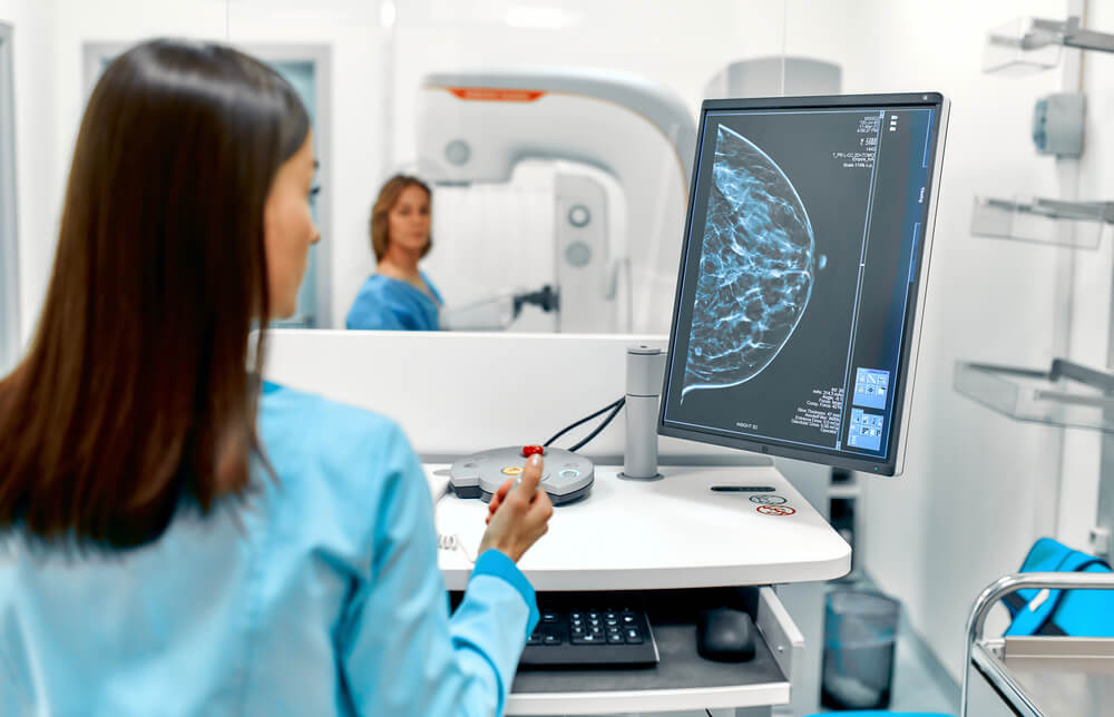
What is Digital Mammography?
Digital Mammography comes to the fore as one of the health checks performed to detect breast cancer. In case of finding a mass in the breast or suspicion, this screening should be performed. We also apply digital mammography within the scope of the screening and tests we perform at our Safir Lab health center. This disease is one of the problems that women encounter quite a lot.
Especially after the age of 40, there may be a case of breast cancer or cyst appearance in women. In case a mass is noticed manually, it is necessary to do this for detailed examination and detection. Our expert medical staff performs digital mammography screening quickly and easily. Within the scope of this imaging that we do digitally, we obtain high quality images. In this way, it can be easily detected in case of cancer.
One of the most common types of cancer in women is breast cancer. Improved screening tests and treatment techniques are saving lives. One of the most common screening methods is a mammogram. It uses X-rays to scan your breasts. The images are checked for any irregularities, and doctors also look for changes in previous tests. The footage was recorded on film for many years. But now digital mammograms can store and analyze information using a computer.
Most people have seen the ads on TV, heard about them on the radio, or come across a magazine article about mammograms. The importance of getting someone done, and the benefits and peace of mind that come with it, are all pointed out. But many people don’t really Decipher the difference between digital mammograms and more traditional movie mammograms.
Simply put, any type of mammogram is a form of X-ray of breast tissue. The main difference is that the old systems were based on film, and digital images were displayed on a computer. Digital mammograms are also easier to view and process. The radiologist can highlight areas, zoom in and zoom out, and in many cases use special filters to see the breast more accurately.
We also offer three-dimensional mammography or tomosynthesis for the early detection of breast cancer and the identification of breast cancer in women with dense breasts.
3-D mammography is a revolutionary screening and diagnostic tool that captures images of the breast in thin sections.
During the procedure, the X-ray arm sweeps the breast and captures a large number of images in a matter of seconds. 3-D imaging allows the radiologist to see the inside of the breast layer by layer, improves fine details by minimizing overlapping tissue, and can be performed in conjunction with a traditional 2-D digital mammogram. 3-D mammography offers October additional advantages, including:
- Detects 41 percent more invasive cancers
- Reduces biopsies and callbacks for a second look by 40 percent
- Takes a picture every four seconds
- Approved by the FDA
- Suitable for all women
Advantages of Digital Mammography
Mammography for breast cancer is quite commonly preferred. This procedure has many advantages and is recommended by our specialist physicians. In case there are any problems with the breasts, this does not appear at once. It will appear over time.
Since breast cancer occurs gradually, it is necessary not to act negligently in this regard. Early diagnosis is very important in breast cancer. If you have breast cancer, you can detect it by having a digital mammography screening at our health center. With the process we have done, we can have detailed information about the audience.
You can have this procedure performed at our health center to find out the size and how well it is. Thanks to the detection of the disease and the mass condition, the possibility of early treatment also arises. Early treatment gives patients a great advantage to get rid of the problem of breast cancer. You can get services from our health center for this.
The advantages of the imaging procedure we perform in our health center are as follows:
- Mammograms printed on film required hard files that were stored with your medical history. A digital mammogram is stored on a computer and can be easily shared electronically, which allows providers to share the necessary data to improve your care.
- A digital mammogram uses ¼ less radiation than its older counterpart. Although earlier doses have been proven to do no harm, less radiation is usually better.
- Finally, doctors and radiologists can make more observations due to better picture clarity and the ability to change photos. Our outstanding radiologists can brighten, darken and enlarge these images almost like an app on your phone. This can make the detection of abnormal changes much more accurate due to the detail provided.
- It offers the possibility of further analysis. Since digital mammograms are stored electronically, they can be analyzed by computers as well as by radiologists.
- It allows for easier second opinions. They can be easily sent electronically for analysis.
- It is useful to see more. Images can be changed for better clarity and visibility. It can’t be a movie mammogram.
Patients should understand that due to the nature of the test, digital mammograms can create a few more false positives.
This means that the scan will pick up even very small changes and differences in the texture that may or may not be relevant. This is known as specificity. Having a high specificity means that more things can be plugged into the network. Specificity is always a balancing act, because it obviously takes more, but not all of it actually matters.
Although mammography captures many small things that are not really important, it also captures small changes that can be early signs of breast cancer. As with any cancer, the sooner it is caught, the better.
As a general rule, digital mammograms are recommended for patients with denser breast tissue. It may also be superior in women who have not yet entered menopause or are under the age of 50.
What is Tomosynthesis Mammography?
Tomosynthesis mammography is a three-dimensional imaging technique performed using X-rays. There is a low dose in this technique that we apply as Safir Lab. It comes to the fore with the fact that it is a procedure applied to many women in our health center. Women can sometimes ignore breast problems or not care enough about these problems. In this case, it is inevitable that the problems will become more serious and important.
In order for me to experience this, even if even the slightest mass is noticed, it is important that you get support from expert people. Masses that appear small at first grow over time. This condition, on the other hand, causes the formation of malignant cysts in women. The biggest reason for the emergence of this situation is negligence. Tomosynthesis mammography performed within the scope of digital mammography, on the other hand, works to detect even very small masses.
We use this method in our health center to examine masses whose size cannot be determined during manual examination or is not visible to the eye. Thanks to its 3D feature, it also provides easier diagnosis. By using these devices, which have become more useful with the development of technology, we offer effective solutions for diagnosis and treatment in our health center.
How is Tomosynthesis Digital Mammography Performed?
The images we obtain with the digital imaging technique are archived. In this direction, it is also used whenever it is needed. At the same time, our specialist doctors can examine these images in more detail. In case of any suspicion about breast cancer, our doctors may order tomosynthesis digital mammography. Using this imaging technique, we determine how dense the mass in the breast is. Our specialist physicians are offered the opportunity to examine the condition of the mass. At the same time, we complete this Decoupling process in 15 to 20 minutes.
While having a tomosynthesis mammography procedure in our health center, you should not have a metal item or product on you. In the case of metal, there may be a distortion of the images taken. At the same time, the procedure needs to be performed a week after the menstrual cycle.
In addition, it is also important not to use deodorant, perfume and similar things on the day of shooting. During the procedure, the nozzle is fixed by placing it between the two layers Decently. In this way, imaging of the breast with the help of a beam tube is provided. Shooting is done from all sizes. As Safir Lab, we do tomosynthesis mammography in this way.
Digital Mammography Prices 2025
One of the most common health problems faced by women is digital mammogram. With this screening process, both breast cancer can be diagnosed early and treated more easily. The screening method, which allows detailed examination of the breast, is recommended by many specialist physicians. The high resolution images obtained after the scan are transferred to the computer and evaluated by our specialist physicians. Many women prefer this method in order to detect signs of breast cancer. Digital mammography, which is very advantageous with its differences between digital imaging applications, is applied in our health center Dec.
Having the correct diagnosis is a critical first step in providing the best possible treatment. If a screening mammogram detects a concern or abnormality, you will be asked to come in for a diagnostic mammogram. Digital mammograms, breast tissue changes before you or your doctor feel more accurately because they have the ability to show early screening and diagnosis of breast cancer plays a critical role in. Our state-of-the-art digital mammogram equipment in our health center allows doctors to view images in real time and examine the breast in more detail.
The method of taking a mammography image is the same for both types. First, your chest is placed between two plates, and then it compresses Decently. They then take top-to-bottom and side-to-side images of your chest. It can be uncomfortable, but the whole process takes about 20 minutes. Film mammograms are recorded in stationary files. With the digital type, X-rays are converted into electrical signals that can be stored on a computer. It is similar to the way digital cameras take and store photos.
In the field of health, each hospital and health center has its own price policy. In other words, pricing is made specifically according to the health center. There are changes in prices according to the periods. The operations we perform within the scope of digital imaging techniques have many advantages. In this way, it also plays an important role for human health.
Mammography continues to be the gold standard for screening for early-stage breast cancer. At our health center, we use a digital mammogram to allow radiologists to capture and check images so that abnormalities can be seen more easily. The digital mammogram procedure is basically performed in the same way as a standard mammogram.
Unlike film-based mammography, a digital mammogram uses computer-based electronic conductors to display a picture of the inside of the breast for the clearest, most accurate images to lead to the correct diagnosis. Our radiologists examine the images and discuss the results with you. If the doctor feels that you need any October evaluation, treatment or biopsy, we will try to schedule it on the same day or within a week. Ultrasound and MRI devices are also available for the evaluation of suspicious masses.
If there is a suspicion of breast cancer, you should definitely consult with specialists. Otherwise, your problem may become even bigger. You can have a digital mammography session by making an appointment at our Safir Lab health center. You can contact us immediately to get information about digital mammography prices 2025.