 Whatsapp
 Call Us


Digital Angiography
What is Digital Angiography?
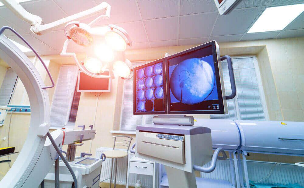
Digital Angiography is a fluoroscopic technique that uses complex X-Ray machines to provide clear images of the inner surface of blood vessels, also called lumens. This technique provides a clear picture of the arteries, veins, and also the four chambers of the heart. This technique is widely used in interventional radiology, as it helps to eliminate radiopaque structures such as bones and provides an accurate image of the blood vessels in question.
Digital angiography is performed to examine and see the vessels in the human body. As Safir Lab, we ensure that all the vessels in your body can be visualized with this procedure that we do in our health center.
Thanks to the examination of your veins, a diagnosis is also made for diseases. This operation is performed within the scope of digital imaging.
In this way, there is no loss of time and the obtained images are viewed from the monitor screen. Within the scope of digital angiography areas such as the brain, legs, abdomen, arms are clearly examined.
During the digital subtraction angiography, the person lies on a table. Electrodes are placed on the chest to monitor heart health throughout the procedure.
An X-ray of the area is taken before the dye is injected. Then, a local anesthetic is injected into the skin in the area where the dye will be applied. Then a thin tube is inserted into the blood vessel and the dye is injected.
October X-ray pictures are then taken, which show the dye in the blood vessels. The computer almost instantly subtracts the structures in the first picture from those in the second picture, leaving behind an orderly picture of the blood vessels of interest.
Without any October procedures, DSA can take as little as 30 minutes. If October additional procedures are needed, the duration may be extended to 3 hours.
What is Digital Angiography?
Veins can be examined for the purpose of diagnosing diseases. This method is used to image every vein in the body.
It is highly preferred in our health center because it provides the clearest images to be obtained. In this way, we diagnose diseases in a short time and determine the appropriate method for treatment.
However, if there is stenosis in the veins, their opening also comes to the fore as one of the methods we apply for diagnosis and treatment.
Digital subtraction angiography (DSA) is a diagnostic procedure that allows a doctor to visualize blood vessels using a contrast medium and digital computer imaging.
The procedure is performed using a catheter, which is a small, flexible, narrow tube that is inserted into an artery in the leg and allowed to travel up to the blood vessels in the brain.
Then, a contrast-forming substance (typically a high-density clear liquid) is injected into the catheter to provide a clear visualization of the blood vessels in the legs, heart, or other organs.
A picture is taken before and after the contrast dye is injected; then the first image is subtracted from the second image to highlight the blood vessels. That is why the method is called Digital Subtraction Angiography (DSA).
How is Digital Angiography Performed?
In the method we apply to view the veins, an entrance is made to the veins using a thin catheter.
You can stay awake while this process is being performed, or if you want, the procedure can also be performed while sleeping. Thanks to the contrast agent we use, we make an examination of even the thinnest of your veins.
In addition, it is also a harmless application to health with minimal use of radiation. This is how digital angiography is performed.
Before performing digital subtraction angiography, the doctor will examine the patient on a number of factors and conditions. Some evaluation parameters include, but are not limited to, the following:
- Any atherosclerotic disease, such as myocardial infection
- Diabetes mellitus
- Kidney function
- Ongoing medications
- Allergies
- Previous surgeries, especially vascular ones
- Historical angiography reports, if any
- Reports of vascular imaging studies related to the procedure
- Any other health condition that could potentially cause complications
After the preliminary evaluation is performed properly, the patient is asked to lie down on the angiography table and local anesthesia is performed.
In some cases, such as if the patient is a child, general anesthesia may be used. Once done, a small incision is made in the leg to insert the catheter into the artery in the leg.
Once inserted, a contrast agent – a dye – is injected into the catheter to produce clear images of blood vessels without any overlapping tissue.
Before the contrast agent is injected into the catheter, a mask image of the corresponding area is taken, which typically shows results just like a normal X-Ray image.
After the contrast agent has been successfully inserted, contrast images are taken through complex X-Ray machines during the time that the contrast agent flows into the artery.
These images are then stored and worked on digitally by subtracting non-contrast (mask images) pixel by pixel to show only the filled veins.
These images can be monitored in real time as they are constantly displayed on the monitor while the examination is ongoing.
After the procedure is completed, hemostasis is performed to the incision site and postoperative care is recommended by the doctor on a case-by-case basis.
Areas of Use of Digital Angiography
Digital angiography, which we perform as Safir Lab, can be used for many purposes. Because it is done for the purpose of examining all veins.
In this direction, it is also quite extensive. In general, the areas of use can be shown in the form of legs, arms and abdominal areas.
In addition, it is also used to control other areas of the body where there are any problems. The skin also presents clear images in areas such as the brain.
Angiography is a technique for producing pictures (angiograms) of internal structures by introducing dye (contrast agent) into the circulatory system, which appears on the X-ray film when the tissue is exposed to x-rays.
The difference between DSA and conventional angiography is that two digital photographs of the tissue are taken with Dec. The first is done before the paint is applied.
The latter is done after the dye is injected. A computer then removes or subtracts certain structures from the first picture from those in the second picture.
This leaves an angiogram that shows only the blood vessels and omits the surrounding or background tissues.
DSA is used to detect blood clots, tumors and other blockages in blood vessels and some ducts.
The procedure is also used to examine the health of blood vessels after coronary artery bypass or other vascular grafting operations.
Sometimes other procedures are performed during a DSA, such as inflating a small balloon in an artery to open the blockage (angioplasty), inserting a stent or metal mesh to keep an artery open, opening a blocked bile duct or duct, inserting a drainage tube, etc.
It is also useful in the operations of blocking the renal canal and inserting a pacemaker device into the chest. During this time, the image guides the physician performing the procedure.
Digital Subtraction Angiography is used to diagnose various diseases related to blood vessels. In order to control occlusive vascular diseases caused by blockage or narrowing in the inner part of the arteries and veins, a person may be recommended to have digital subtraction angiography.
In addition, the procedure can also be used to diagnose other conditions, such as a brain aneurysm, bleeding vessels, unusual artery and vein connections, and to analyze the blood vessels of a tumor.
However, the technique also helps with various interventional procedures, as it helps to achieve improved visibility. Some conditions in which the technique can effectively help are:
- Endovascular aneurysm repair
- Arterial balloon angioplasty
- Arterial stent
- Endovascular embolization
- Thrombectomy
- Nephrostomy
- Biliary procedures
Coronary Angiography
Since veins are all over the body, they are used to detect different problems. Coronary angiography is a method performed in this context and used to examine the heart vessels.
The procedure is performed under local anesthesia and lasts an average of 15 minutes. We make an entrance through the groin area using a thin catheter. In this direction, we also view the vessels that feed the heart. At the same time, you will be under observation for 2-6 hours after the procedure is completed.
Micro Angiography
The technique used for imaging capillaries is called micro angiography. In this method, we do the procedure much more carefully than according to the image of the arteries and veins. In this imaging technique, we use the thinnest catheter.
Local anesthesia is used in order for the patient to have no pain or to have a minimal amount. With this method, which is among the digital angiography methods, it is possible to obtain clear images of the capillaries Decisively.
Neurovascular Angiography
The method we use to examine the vessels leading to the brain is called neurovascular angiography. By performing this procedure in our health center, we ensure that both the arteries and the blood vessels leading to the brain are clearly visualized.
In general, it is a frequently preferred way of blood flow assessment. We perform procedures for our patients under local anesthesia. In this way, we prevent you from experiencing pain or pain. If pain occurs after angiography, it can be easily relieved with medication.
Peripheral Angiography
The angiography method used in the problem of vascular occlusion is called peripheral angiography. We use this method in our health center for the opening of vascular occlusion.
The reason why congestion occurs is that there is little blood flow to the feet and legs. It is one of the digital angiography methods that we apply in our health center in order to find out what causes this blockage.
Can Digital Angiography Be Performed for Every Patient?
Contrast agent is given from the vein in angiography applications performed in our health center. In this context, the veins can be examined.
This is the most commonly used method to clearly see the coronary vessels. This procedure can be easily performed for each patient.
Our specialist physicians and medical personnel will help you by performing the procedure in a practical way. It does not threaten the patient’s health in any way and is extremely reliable.
A DSA is a relatively safe procedure with very rare complications such as:
- Drainage or bleeding from the incision site
- Formation of a pseudoaneurysm at the puncture site
- The harmful effect of contrast medium on other organs, such as the kidneys
- Hypersensitivity or allergy to contrast medium
In general, a digital subtraction angiography is a safe, easy, fast and cost-effective procedure that can also be performed on an outpatient basis to obtain information that would not otherwise be obtained from a conventional angiography.
In addition, with technological progress, digital subtraction angiography provides greater contrast sensitivity and therefore constitutes a very powerful alternative to conventional angiography. The results of the procedure can be obtained immediately, and besides, they are very accurate, which adds even more reliability to the method.
Digital Angiography Prices 2025
Heart and vascular diseases are among the important health problems. Dec. To diagnose these diseases, imaging of the vessels is required. We also perform this procedure with angiography performed at our health center. The prices of the procedure vary according to the hospital and health center where the service is received.
A DSA should provide a clear picture of the blood vessels of interest. If the picture is insufficient to make a definitive diagnosis, other tests may be performed. These include a conventional (non-removable) angiogram, duplex sonography or a magnetic resonance angiogram (MRA).
After DSA, a person needs to stay still for about 6 hours to prevent bleeding. During this time, the person will be monitored for complications. Depending on the appearance of complications, the person’s health and the October procedures performed, it may be necessary to stay in the hospital overnight. You can get information about digital angiography prices and make an appointment by contacting our Safir Lab health center. You can contact us immediately to get information about digital angiography prices 2025.