 Whatsapp
 Call Us


Chest X-Ray
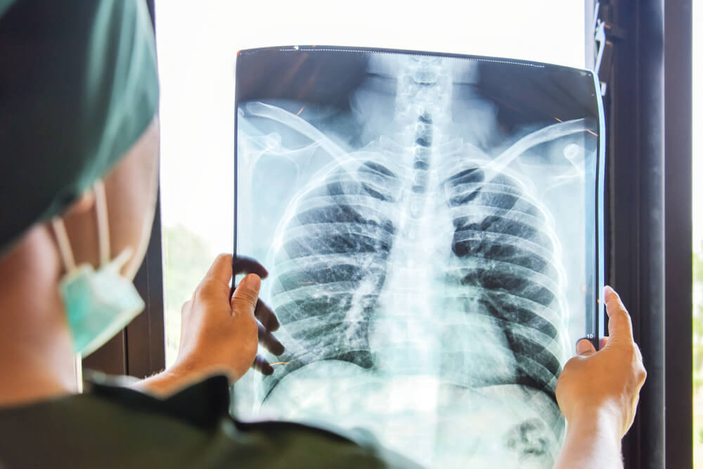
What is a Chest X-Ray?
Chest X-ray is performed to obtain detailed information about the lungs. It is useful to perform this procedure so that the chest area can be examined in a better way. As one of the imaging techniques, it is preferred in diagnosing people’s diseases and monitoring their health status.
We perform this procedure with our specialist doctors at our Safir Lab health center. While this application is beneficial for human health, it allows doctors to diagnose diseases in a more comfortable way.
Especially lung disorders, if neglected, can turn into serious problems. That is why it is extremely useful for your health to have a lung X-ray at our health center. Lung diseases may indicate cancer in some cases. If this progresses, vital problems may arise. Our doctors may ask you to have a chest X-ray for this condition if it is deemed necessary.
The procedure performed on the chest X-ray is twofold. The first stage is called step graphy. Later, an examination is performed to diagnose the disorders. Imaging of the lungs is performed with frontal and lateral shots. In case of problems in the lungs, heart and similar organs, it can be learned in this way.
A chest X-ray is a test that creates an image of your heart, lungs, and bones. Another name for a chest X-ray is a chest radiography.
X-rays use focused beams of radiation. These radiation rays create pictures of the inside of your body. X-ray images are similar to negative images of black and white photographs.
A chest X-ray is a very useful examination, but it has its limitations. Since some conditions of the chest cannot be detected on a conventional lung X-ray image, this examination cannot necessarily rule out all problems in the chest. For example, small cancers may not be visible on a chest X-ray. A blood clot in the lungs, a condition called pulmonary embolism, cannot be seen on chest x-rays.
Further imaging studies may be necessary to clarify the chest X-ray results or to look for abnormalities that are not visible on the chest x-ray.
Why is a Chest X-Ray Requested?
Radiography may be requested to diagnose all kinds of health problems that affect the lungs or may be caused by the lungs. It is also effective in detecting many breast diseases. The treatment process can be started by detecting the health problems indicated by chest X-ray. You can benefit from our services by having an X-ray taken at our health center.
A chest X-ray helps doctors at our health center diagnose problems in your heart or lungs that are causing symptoms. Some of these symptoms are:
- Chest pain.
- Chronic cough.
- Difficulty breathing.
- Fever with other signs of infection.
Your healthcare provider may also recommend a chest X-ray to diagnose or monitor certain health conditions, including:
- Congestive heart failure.
- Emphysema or chronic obstructive pulmonary disease (COPD).
- Lung cancer.
- Pneumonia.
- Injuries to the rib cage.
Always tell your healthcare provider if there is a possibility that you are pregnant. Exposure to radiation can harm a developing baby. In general, the amount of radiation used for simple chest X-ray is so small that it is considered safe during pregnancy. However, our healthcare professionals will help you make the decision to have an X-ray based on the urgency of your symptoms.
X-rays use a very small amount of radiation. For adults, the risks are minimal. In younger children, lower radiation X-rays can be used to minimize the risk in this population.
Radiography is requested by our doctors for people who show symptoms. This is how the detection of the disease is ensured. For the detection and treatment of all problems related to your lungs, you can easily have a chest X-ray at our Safir Lab health center.
How Is the Lung X-Ray Preparation?
It is important that you inform our doctors about some issues before taking a chest X-ray for your lungs. First of all, if you have a condition such as pregnancy, you should definitely share it. As a result of a chest X-ray, a low radiation is taken. In this direction, radiography is not a harmful or a procedure that should be feared.
It is extremely important for people suffering from respiratory distress to have a radiograph in terms of health. Our expert medical personnel inform you about the requirements to be followed before the procedure.
A chest X-ray requires little or no preparation. When preparing for an appointment, wear loose, comfortable clothes that do not contain metal (zipper, studs, bra closure) and leave your jewelry at home. If you have a body piercing, consult our health center for specific instructions. Body jewelry can interfere with clear images. You may need to remove it or replace it with an acrylic holder.
Occupational Groups That Have to Undergo Lung Radiography
Lung diseases can be shown as a disease that covers all business sectors. It is especially important for people working in factories in the industrial sector to have their lungs checked. It is possible to examine the condition of the lungs with a chest X-ray, which will be performed once a year. You can have this procedure done easily at our health center. Our lung specialist doctors or radiology specialists help you with this.
This process that we have done comes to the fore with the fact that it is both safe and shows the right results. You can get services from our health center for the diagnosis of lung diseases. The lungs, located on the right and left sides in the chest cavity, can be checked by chest X-ray and X-ray method.
Having a film of your lungs taken will be very useful for you. The advantages of this process can be shown as follows.
- It is not the case that radiation remains in the body of people after X-rays are taken.
- X-rays usually have no side effects within the typical diagnostic December for this examination.
- X-ray equipment is relatively inexpensive and is widely available in emergency rooms, doctor’s offices, outpatient centers, nursing homes and other places, which makes it convenient for both patients and doctors.
- Since X-ray imaging is quick and easy, it is especially useful for emergency diagnosis and treatment.
Disorders Detected By Chest X-Ray
Having a radiograph plays a role in detecting many problems related to your lungs. In this way, health problems can be detected early and treatment can be started immediately. It is very important to take a chest X-ray, especially for smoking-related problems. In this direction, you can find out the condition of your lungs.
In case of damage to the lung tissue, it is also necessary to undergo a chest X-ray to diagnose it. We help you by detecting all kinds of lung diseases in our health center.
Radiography can also be performed to follow up the detected nodules. Nodules may not cause a tumor, but it is still important for health to follow up. Tissue inflammation, you can also have a radiograph to determine the negativities in bone structures. We carry out this procedure in our Safir Lab health center.
A chest X-ray helps healthcare providers diagnose problems in your heart or lungs that are causing symptoms. Some of these symptoms are:
- Chest pain.
- Chronic cough.
- Difficulty breathing.
- Fever with other signs of infection.
Our healthcare professionals may also recommend a chest X-ray to diagnose or monitor certain health conditions, including:
- Congestive heart failure.
- COPD or emphysema disease
- Lung cancer.
- Pneumonia.
- Injuries to the rib cage.
Does a Chest X-ray Give Immediate Results?
The radiography method applied for the lungs is an imaging technique that allows immediate results to be obtained. At the same time, he is also so reliable. If an advanced examination is required, you can have a chest X-ray at our health center to determine this.
With a chest X-ray, you can easily find out about your lung problems. In the direction of the respiratory tract, the lungs are organs that are in contact with the outside. In this direction, it is also important to check the lungs with chest X-ray and X-ray for health.
Chest X-ray does not require special preparation. During the procedure, you will be asked to take off some of your clothes and put on an apron. Our medical personnel may request that you remove jewelry, removable dental instruments, and any metal objects or clothing that may negatively affect the images to be obtained from the X-ray.
You can wear a medical gown in our doctors’ office. The X-ray technician will also ask you to remove all metals, such as glasses, jewelry, or hairpins.
Typically, your chest X-ray consists of two parts:
- You are standing with your chest resting on the metal plate of the X-ray machine and your hands on your hips. This position creates an image of the front of your chest.
- You are standing with your side to the metal plate of the X-ray machine and your arms in the air. This position creates an image of the side of your chest.
You may be asked to stay very still and hold your breath during a chest X-ray. Any movement, even breathing, can blur the X-ray image. Chest x-rays usually take a few minutes to complete.
After the X-ray, your radiation technician may ask you to wait a few minutes while looking at the images. If any of the images are blurred, the technician may need to take the X-rays again.
X-ray images are sent to a radiologist who examines them for normal and abnormal findings. Our healthcare professionals will then review the images and the radiologist’s report so that they can discuss your X-ray results with you.
Our doctors use the examination to help treat – diagnose or monitor a number of conditions that may occur in humans:
- Pneumonia
- Heart failure and other heart problems
- Emphysema
- Lung cancer
- Positioning of medical devices
- Collection of fluid or air around the lungs
- Other medical conditions
A chest X-ray is a test that looks at your heart, lungs, and bones. A chest X-ray uses a small dose of radiation to create a black and white image. Healthcare providers can look at this picture to diagnose and treat broken bones, heart conditions, and lung problems. Chest X-rays are fast, non-invasive procedures performed by our health center.
Diagnosis of Lung Diseases by Chest X-Ray
In case of a problem with your lungs, this determination is made by our specialist doctors and medical personnel. You should inform us about your medical history so that our doctors can make a good assessment and make a clear diagnosis. At the same time, it is important that you provide the necessary information about your work environment. After learning the necessary information, you can have a chest X-ray at our Safir Lab health center.
We use advanced equipment to diagnose your diseases. If you want to find out if there is a problem with your lungs, you can make an appointment at our health center for this. Then the processes of making a diagnosis and determining the appropriate treatment begin.
A chest X-ray allows you to view the inside of the chest with a small dose of ionizing radiation. It is used to assess the lungs, heart, and chest wall, and can be used to help diagnose shortness of breath, persistent cough, fever, chest pain, or injury.
It can also be used to help diagnose and monitor the treatment of various lung conditions, such as pneumonia, emphysema, and cancer. Since chest X-ray is quick and easy, it is especially useful for emergency diagnosis and treatment
The thickness of the tissues of your body varies. When radiation passes through your body, each structure in your body allows a different amount of radiation to pass through. For example, your bones are very thick and do not allow much radiation to pass through. The bones appear white on the X-ray image.
However, your lungs allow more radiation. Your lungs look gray on an X-ray image. As a healthcare provider, we look at the colors and shades on an X-ray to diagnose and treat health conditions.
Chest X-Ray Prices 2025
You can contact us immediately to get information about lung radiography prices 2025.